Observations:
Histochemistry staining
- Compacted
- Looks like a network
- Multiple layers
- Purple nucleus - due to a single stain
- Pink cytoplasm - again from a single stain
- Both stains do not affect the others. Eg: Purple stain does not stain cytoplasm while pink stain does not stain nucleus
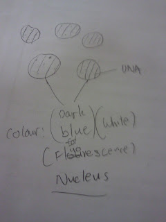
Fluorescence staining
- Stain DNA only (makes nucleus fluorescent due to Hoechst Dye)
- Background is black
- Able to see DNA in nucleus at 100x Magnification
- Organelles w/o DNA are not stained.
Immunohistochemistry staining
- Dark brown cells
- Light brown layers
- Multiple layers
- Have many gaps
- Connected layers
What is the end product/ findings?
- When hematoxylin and eosin was used on the sample, a liver cancer tissue, we found that the hematoxylin stained the nuclei but not the cytoplasm while eosin stained the cytoplasm but not the nuclei.
- Normal cells of the tissue overlap each other neatly while cancerous cells will space out, leaving large spaces in between, and not overlap each other.
- The structure of the fluorescent microscope is totally the opposite of that of the light microscope.
- The Hoescht dye lights up under a specific wavelength of UV light and stains organelles containing DNA, but the background of what is seen is black.
- Arteries are able to be spotted through the microscope between the cells that we see.
A description/ explanation of our product/ findings.
- The nuclei are stained by hematoxylin, in shades of blue to purple, since they have negatively charged DNA, while the hematoxylin is positively charged. On the other hand, eosin is negatively charged and will bind with the positively charged proteins in the cytoplasm of the cell, staining the cytoplasm in shades of pink, orange and red.
- This is as normal cells will work together as a tissue while when they become cancer
cells, they have a mind of their own already, not working as a tissue anymore.
- This is as for some of the samples scientists have to view where the cells are at the bottom of the container, the fluorescent microscope allows a better view of these cells as the light comes from the bottom instead of coming from the top.
- The Hoescht dye is fluorescent, and when it is excited by the certain wavelength of UV light, it appears brightly causing it to stand out from the black background, as there is no light in the background switched on except for the UV light, which only excites the stained nuclei.
- Blood capillaries appear in the form of gaps in between the cells where they are large enough when magnified to be seen through the microscope.
Some links to our pages:
Back to our team...
Back to home...
An overview...
Our mission for this project...
From this project... we have gained...
From this project... we have reflected...


